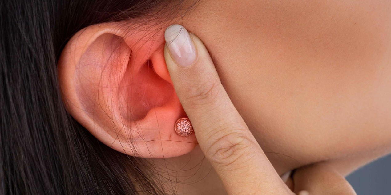
Our ears are the gateways for sound to enter our world; they are like tiny machines that carry both whispers and our laughter. However, one of these complex structures—both the outer ear and the ear auricle—can rarely become a nest for malignant cells. This condition called “ear cancer,” although often beginning as an extension of skin cancers, can lead to serious problems in terms of hearing, beauty, and even life comfort when delayed.
What is Ear Cancer?
Ear cancer, as the name suggests, describes malignant tumors resulting from uncontrolled division of cells in the tissues that make up the ear. It can be examined under two main headings:
External Ear (Ear Auricle and External Canal) Tumors:
- Basal Cell Carcinoma (BCC): The most common, slow-growing skin cancer; appears as fine wounds or pink nodules on the ear auricle.
- Squamous Cell Carcinoma (SCC): More aggressive; can turn into crusty, bleeding lesions or non-healing wounds.
- Melanoma: Manifests as colored or pigmented lesions; can grow rapidly and penetrate deep tissues.
Middle Ear and Inner Ear Tumors (Rare):
- Keratocystic Odontogenic Tumor, Adenoid Cystic Carcinoma: Usually associated with chronic infection or previous radiation history.
- Paraganglioma, Vestibular Schwannoma (Acoustic Neuroma): Actually considered “benign” but can cause hearing and balance problems by pressing on surrounding tissues as they grow.
While external ear cancers can be treated with high success rates through surgical excision when caught early, middle and inner ear tumors require multidisciplinary approaches.
What Causes Ear Cancer? (Risk Factors)
UV Rays and Sun Damage
We must not forget that the ear auricle is one of the most sun-exposed areas. If we don’t use hats or sunscreen, our risk of basal and squamous cell carcinoma increases.
Chronic Infections and Psoriasis
Long-term otitis externa or middle ear inflammations can increase mutation risk by accelerating the cellular repair cycle in the area.
Radiation and Previous Cancer Treatments
Radiotherapy received to the head-neck region can prepare the ground for tumor development in ear skin or deep tissues years later.
Chemical Exposure
Long-term work with arsenic, tar, and some organic solvents in industrial environments increases skin tumor risk.
Fair Skin and Family History
Individuals with low melanocyte count, easily burned skin, or similar cancer history in the family have higher risk.
Immune System Suppression
Medications used after organ transplantation or conditions causing immune weakness like HIV/AIDS can invite both skin and inner ear tumors.
Who is at Risk for Ear Cancer?
- Fair-skinned and Freckled Individuals – Those with severe reaction to sun, who don’t tan.
- Those Working Outdoors for Long Periods – Fishermen, construction workers, gardeners.
- Those Who Previously Received Head-Neck Radiotherapy
- Those with Chronic Ear Infections – Especially those with external ear canal problems.
- Immunocompromised Patients – Organ transplant recipients, HIV positive individuals.
- Individuals Over 50 – DNA repair mechanisms weaken with age.
If you’re in one of these groups, instead of worrying “Do I Have Ear Cancer?”, make adding regular ear examination to your calendar a habit.
Diagnosis and Evaluation Process
Clinical Examination and Otoscopy
Careful visual examination of ear auricle and external canal; examining suspicious area with magnification (dermatoscope).
Dermatoscopy and Photodocumentation
Evaluates the lesion’s color, texture, vascular structures, and borders at microscopic level, records future changes.
Biopsy
- Punch or Incisional Biopsy: In superficial lesions, a small tissue piece is taken under local anesthesia.
- Excisional Biopsy: In larger or deeper lesions, the entire tumor area is removed and sent to pathology.
Imaging Studies
- CT/MRI: Middle and inner ear involvement, bone erosion, or parotid, facial nerve proximity is detected.
- PET-CT: Used to evaluate suspicious metastasis or lymph node involvement.
Multidisciplinary Evaluation
ENT specialist, dermatologist, plastic surgeon, and oncologist come together to determine treatment plan.
Ear Cancer Treatment Methods
Surgical Excision
- Wide Local Excision: In external ear cancers, lesion is removed with 5-10 mm safety margin.
- Parotid and Lymph Node Dissection: Depending on tumor size and spread, intervention to parotid gland or neck lymph nodes may be needed.
Mohs Microsurgery
Layer-by-layer pathology examination providing maximum clearance with minimal tissue loss in external ear; preferred especially in face and ear regions.
Radiotherapy
In elderly patients or those with poor general condition where surgery cannot be performed, or as adjuvant to surgery, providing control in target area with rays instead of closing incision.
Chemotherapy and Immunotherapy
In rare middle ear carcinomas or metastatic squamous cell cancers, platinum-based chemotherapies or anti-PD-1/PD-L1 agents may come into consideration.
Reconstructive Surgery
Local flap, free flap, or ear auricle reconstruction may be needed to provide wound repair in extensive tissue excisions.
Treatment selection is customized considering tumor type, spread, patient’s age, and general health condition.
The Importance of Specialist Selection
Although ear cancer may be perceived as a “small skin lesion,” considering the form of ear auricle and hearing function, it requires meticulous intervention both functionally and aesthetically. An ideal treatment team includes:
- ENT Specialist/Head & Neck Surgeon: Knows the tumor’s effect on hearing and facial nerves.
- Dermatologist: Manages subcutaneous spread risk and recurrence monitoring.
- Plastic Reconstructive Surgeon: Restores ear form to its most natural state after excision.
- Radiation Oncologist and Medical Oncologist: Plan adjuvant treatments.
This multidisciplinary team considers both “eliminating cancer” and “preserving ear integrity” goals together.
What Awaits the Patient on Treatment Day?
- Morning Preparations and Measurements: Blood tests, EKG, nutritional status are reviewed.
- Anesthesia Consultation: Local (usually) or general anesthesia decision is made.
- Surgical Procedure: Duration varies from 30 minutes to several hours; in Mohs surgery, layer-by-layer pathology examination may be divided into several rounds.
- Reconstruction: Flap or graft application after extensive excision.
- Recovery and Observation: Same-day home return after simple excision, 1-2 night hospital stay for extensive interventions.
Pre and post-operative jokes by the team dispel tension; sometimes you might hear the joke “Now I’ll be able to listen to the wind’s messages with my ears too!”
Post-Treatment Recovery and Follow-up Process
- 1-2 Weeks: Sutures are removed in 7-10 days; swelling, mild pain, and sensitivity are normal. Cold compress and prescribed creams increase comfort.
- 1-3 Months: While swelling decreases, flap or graft adaptation and color differences are closely monitored. Using sun-protective hat and SPF 50+ cream prevents pigmentation.
- 6-12 Months: Ear form and sensation take final shape; hearing test checks middle ear function.
- Annual Check-ups: Although recurrence risk is low, regular dermatology and ENT examination is recommended.
Frequently Asked Questions
1. Will there be scarring on the ear auricle?
Depending on surgical technique, a thin line may remain; its prominence decreases over time with subcutaneous tissue renewal.
2. Will my hearing be affected?
External ear auricle and canal level procedures usually don’t affect hearing; hearing tests are performed in middle or inner ear surgery.
3. Is Mohs surgery painful?
It’s almost painless with local anesthesia, performed in sessions lasting several hours.
4. Can I go in the sun after radiotherapy?
High-protection hat and SPF 50+ cream are mandatory until treatment ends; protect from skin phototoxicity.
5. Is there risk of recurrence?
Cure rate is high with early-stage excision and/or Mohs microsurgery; however, annual check-ups help catch new lesions.
Conclusion and Take the First Step
Ear cancer is a condition that goes far beyond the perception of “a small crust,” directly related to hearing and appearance. Early diagnosis, multidisciplinary treatment with specialist team, and disciplined follow-up are the most powerful ways to eliminate this threat. Now, don’t ignore any changes in your ears; make an examination appointment with your ENT specialist or dermatologist. Because your ears are not only valuable guides for sound, but also for your health!

