Our face is the most intimate stage where we tell the story of our life: every smile, every thought leaves its mark. However, this delicate and visible area is also most vulnerable to the sun, harmful UV rays, and sometimes genetic misfortune—making it a host for a serious threat called “facial skin cancer.” Facial skin cancer can be controlled with early diagnosis and the right treatment approaches, but when neglected, it may pose life-threatening risks.
What Is Facial Skin Cancer?
Facial skin cancer refers to malignant tumors resulting from uncontrolled cell division in the skin of the face. Skin cancers are generally divided into three main categories:
- Basal Cell Carcinoma (BCC)
The most common (over 80%) and slowest-growing type. Typically appears on sun-exposed areas such as the forehead, nose, and cheeks. It manifests as pearl-like nodules or open sores. - Squamous Cell Carcinoma (SCC)
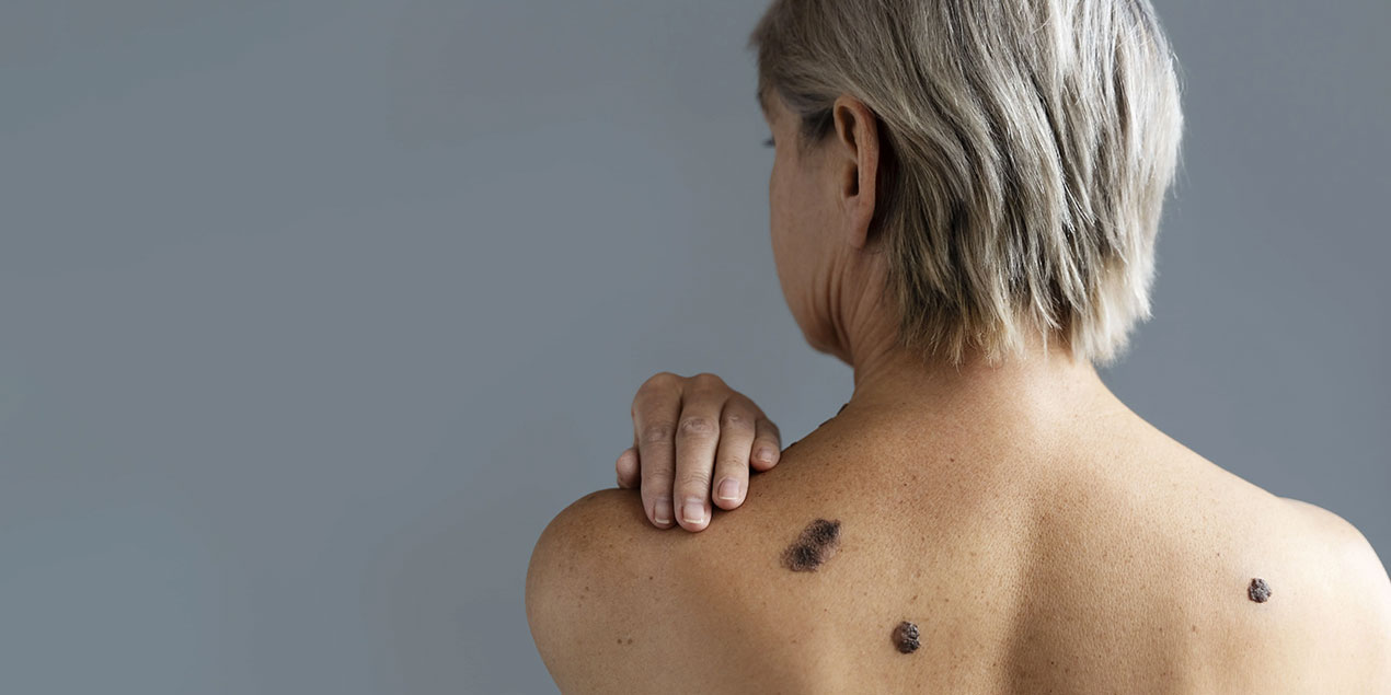
More aggressive and deeper spreading than basal cell carcinoma. It may present as red, crusted lesions or non-healing wounds. - Melanoma
Considered the most dangerous type; starts as a pigmented (brown-black) spot or mole and can rapidly invade deep tissues. If not detected early, it carries a high risk of metastasis.
Although the term “skin cancer” may provoke fear, early diagnosis followed by surgical and medical interventions can offer a high cure rate. The key is to take that “minor flaw” or persistent scab seriously and consult your dermatologist.
What Causes Facial Skin Cancer? (Risk Factors)
1. Ultraviolet (UV) Exposure
The gentle rays of the sun unfortunately aren’t friendly to our skin. UV-A and UV-B rays cause DNA damage in skin cells, increasing cancer risk. Don’t skip the hat or high-SPF sunscreen at the beach!
2. Tanning Beds and Sunlamps
Tanning, often seen as the “fast track to a golden glow,” bombards your skin with UV rays. It significantly increases the risk of both premature aging and skin cancer.
3. Fair Skin and Sun Sensitivity
People with low melanin (fair skin) have a weaker protective barrier. If you have a history of sunburn, you need to be extra cautious about skin cancer.
4. Advanced Age
As we age, our DNA repair mechanisms slow down, and skin cells that have accumulated UV damage become riskier. However, high sun exposure and tanning in youth are shifting these statistics.
5. Genetic Predisposition and Family History
If skin cancer runs in your family, your risk increases. Use of synthetic creams, certain medications (e.g., chemotherapy agents), and immunosuppressive conditions are also risk factors.
Who Is at Risk for Facial Skin Cancer?
- Fair-skinned, freckled individuals who do not tan easily
- Those with high sun exposure (outdoor sports, beachgoers)
- Tanning bed users
- Immunosuppressed individuals (e.g., organ transplant, HIV/AIDS)
- People over age 50
- Those with a family history of melanoma syndrome
If you fall into one of these groups, don’t panic—just be sure to have regular dermatology checkups and get suspicious lesions examined promptly.
Diagnosis and Evaluation Process
1. Clinical Examination
Your dermatologist will carefully examine your skin, assessing the lesion’s color, shape, size, border, and any history of change. The ABCDE guide is used with questions like “Is it asymmetrical?”, “Are the borders irregular?”, “Is the color uniform?”:
- A (Asymmetry)
- B (Border irregularity)
- C (Color variation)
- D (Diameter greater than 6 mm)
- E (Evolution – rapid changes)
2. Dermoscopy
A handheld magnifying tool is used to examine the lesion in depth. Vascular structures, melanin patterns, and microscopic features are evaluated to better identify suspicious tissue.
3. Biopsy
A definitive diagnosis requires a tissue sample (punch, excisional, or incisional biopsy). Pathology reveals the type and stage of cancer. It might feel like a “mini surgery,” but local anesthesia ensures your comfort.
4. Imaging (if necessary)
For suspected melanoma or invasive squamous cell carcinoma, ultrasound, CT, or PET-CT may be used to check lymph nodes and metastasis.
The greatest advantage of this stage is to “break the tumor” early and allow for complete planning—not just “break a leg.”
Treatment Options for Facial Skin Cancer
1. Surgical Excision
The lesion is removed along with a margin of healthy tissue (typically 3–5 mm). For the aesthetically critical facial area, tissue-sparing techniques are used to achieve both full cure and good cosmetic results.
2. Mohs Micrographic Surgery
This precise method removes the cancer layer by layer, with pathology evaluation after each layer. It offers the highest cure rate (up to 99%) and minimal tissue loss—especially preferred for facial cancers.
3. Radiotherapy
Used when surgery is not an option (elderly or comorbid patients), or as an adjunct therapy. Side effects include local redness, dryness, and rarely, pigment changes.
4. Topical Treatments
In early-stage superficial basal cell carcinoma or precancerous lesions (actinic keratosis), creams like imiquimod or 5-fluorouracil may be used. Consistent application and close follow-up are essential.
5. Systemic Therapy and Immunotherapy
For advanced melanoma or metastatic squamous cell carcinoma, anti-PD-1/PD-L1 therapies (e.g., pembrolizumab, nivolumab) or targeted therapies (e.g., vemurafenib, dabrafenib) are used. These activate the immune system or block cancer growth signals.
Treatment is selected based on cancer type, size, depth, patient age, and overall health, through a multidisciplinary evaluation by oncology, dermatology, and plastic surgery teams.
The Importance of Specialist Selection
The facial skin is both a functional and aesthetic focal point. Treatment should aim not only to eliminate cancer but also ensure optimal cosmetic outcomes. The ideal approach includes:
- Experienced Dermatologist/Plastic Surgeon: Proficient in Mohs surgery and oncoplastic reconstruction
- Multidisciplinary Team: Collaborative work between oncology, radiology, and pathology specialists
- Advanced Technology and Equipment: Dermoscopy, digital pathology, modern radiotherapy tools
- Empathy and Communication: A physician who transparently explains the process and listens to your concerns
These four factors ensure maximum success with minimal scarring on your face.
What to Expect on Treatment Day?
- Preparation and Consultation: Blood tests and anesthesia consultation (local anesthesia is common for surgery)
- Procedure Duration: Simple excisions take 30–60 minutes; Mohs surgery may last 2–4 hours (due to layer-by-layer evaluation)
- Comfort Measures: Pain is minimized with anesthesia; sedation may be given if necessary
- Skin Repair: Post-excision closure typically involves flaps, grafts, or primary sutures
- Discharge: Most patients go home the same day; rarely, an overnight stay is needed
Recovery and Follow-Up Process
- First 1–2 Weeks: Sutures are removed in 7–10 days. Mild swelling and sensitivity are normal. Cold compress and prescribed creams help comfort.
- 1–3 Months: Scar gradually improves; wearing sun-protective clothing and SPF 50+ creams reduces pigmentation risk
- 6–12 Months: The scar softens and blends closely with your skin tone. Regular checkups at 3, 6, and 12 months are recommended
- Annual Screenings: Yearly visits to a dermatologist are crucial for detecting new lesions early
Frequently Asked Questions
1. Will there be a scar?
Depending on the surgical technique and lesion location, a thin scar may remain but usually fades over time.
2. Does Mohs surgery take a long time?
Yes, it involves step-by-step pathology review; typically 2–4 hours—but it offers the highest cure rate.
3. Is radiotherapy painful?
Pain during treatment is rare; mild redness and dryness afterward are common.
4. How can I detect new lesions?
Perform monthly skin self-exams using the ABCDE guide; any rapidly changing or non-healing wounds should be evaluated.
Conclusion: Take the First Step
Facial skin cancer is more than “just a small spot”—it’s a threat to both your health and aesthetic integrity. With early detection, the right specialist, and disciplined follow-up, it can be effectively managed. Now is the time to approach every mark in the mirror with awareness—not fear. Schedule an exam with a dermatologist or oncoplastic surgeon today and take the first step toward protecting your face and quality of life!